Identification
Mushroom Bruising and Color Changes Explained
Document mushroom bruising by location, trigger, timing, and color while separating oxidation from age, spores, latex, and damage.
Safety first
Use as a field-record guide
Document the whole mushroom, habitat, substrate, and region before comparing names or photographs.
Open the identification safety hub →Decision limit
Do not use this as an edibility verdict
Wild mushroom consumption decisions require qualified local, in-person confirmation of the complete specimen.
Read the safety disclaimer →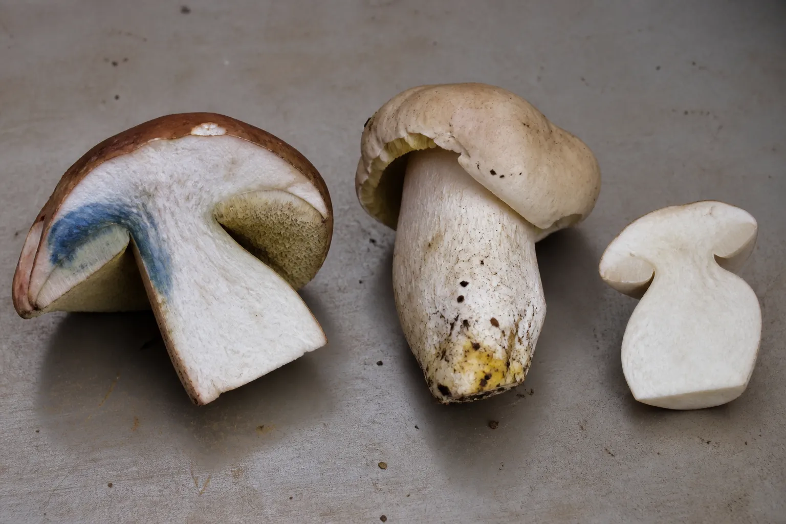
Safety scope: This editorial guide explains evidence and documentation. It cannot confirm that a wild mushroom is edible, and it has no named expert or clinical reviewer.
What mushroom bruising actually records
Bruising is a color response after tissue is pressed, cut, or otherwise damaged. It often involves enzymatic oxidation of compounds exposed when cells rupture, but chemistry differs among fungal groups. Record the original tissue color, exact contact point, delay before change, maximum color, and later fading. A reaction is one character in a larger identification, never a verdict.
Darkening from age, waterlogging, frost, bacterial decay, or drying is not equivalent to a fresh bruise. Spore deposits can color caps below a cluster. Milkcap latex may change after exposure while the flesh behaves differently. A scraped cap surface may merely reveal pigment below the cuticle. State which tissue changed rather than saying only that the mushroom “turned blue.” For the complete evidence sequence, use the identification safety checklist.
Bruising is best treated as a timed observation. The useful record is not only the final color, but the intact starting color, trigger, anatomical location, delay, direction of spread, maximum intensity, and later fading or darkening. Those variables can separate patterns that look identical in a single finished photograph.
Standardize pressure and cut observations
Use one uninjured specimen as a control. Apply gentle pressure to a defined cap or pore area and make one clean cut through a second section. Photograph immediately and at consistent intervals in neutral light. Aggressive squeezing can mix soil, spores, and damaged layers, producing a result that another observer cannot reproduce.
Choose a fresh, representative specimen and leave one comparable area untouched as a control. With clean tools, apply one gentle press to a defined cap, pore, gill, or stem location and make one clean lengthwise cut. Avoid crushing the whole specimen: pressure can mix layers, soil, spores, and fluids into a color that cannot be reproduced.
Photograph the intact surface, immediately after injury, then at fixed intervals such as 30 seconds, two minutes, ten minutes, and thirty minutes. Slow reactions may justify a later frame. Keep light, white balance, angle, and exposure consistent, and include a scale and neutral color reference. The mushroom photo checklist shows how to preserve anatomical context.
Record ambient temperature and whether tissue is wet, dry, chilled, old, or waterlogged. Do not compare a refrigerated market specimen with a fresh field specimen as though reaction speed were controlled.
Separate bruising from lookalike changes
Darkening from age, waterlogging, frost, bacterial decay, or drying is not equivalent to a fresh bruise. Spore deposits can color caps below a cluster. Milkcap latex may change after exposure while the flesh behaves differently. A scraped cap surface may reveal underlying pigment without a chemical reaction. State which tissue changed rather than saying only that the mushroom turned blue.
First ask whether the color begins exactly where fresh tissue was injured. Diffuse darkening already present before handling may reflect age or decay. A colored layer revealed by scraping is exposure, not necessarily a chemical change. Soil can stain a stem base; rusty or dark spores can dust lower caps in a cluster; colored latex can spread from cut gills into nearby flesh.
Use paired views of intact and injured tissue. Note whether cap flesh, pore layer, tubes, gills, stem cortex, stem base, or latex changes independently. “The mushroom bruises” discards the anatomical detail needed by a key.
Odor, spore deposit, substrate, host trees, complete base, maturity, and microscopic characters remain separate evidence. A convincing bruise reaction does not compensate for a missing base or unknown substrate.
How blue, yellow, and green reactions differ
Many boletes show blue reactions after pores, tubes, or flesh are damaged, but the location and tempo vary. One collection may blue instantly in the pores while context flesh changes slowly; another may show little reaction in old or dry material. Both toxic and edible taxa occur among blue-staining boletes, so blue is neither a poison alarm nor an edibility certificate. Use the bolete identification guide to place staining beside pore color, stem ornament, cap, context, habitat, and regional taxonomy.
Some Agaricus show yellow after rubbing the cap margin or cutting the stem base. The intensity, persistence, exact site, odor, and temperature can matter in a regional key. A weak yellow cast in an aged photograph is not equivalent to a prompt basal reaction observed in person.
Milkcap records require two linked observations: the initial latex color and any later change in the latex or damaged flesh. White latex that yellows, orange latex that becomes greenish, and flesh that stains independently are different descriptions. Latex quantity also changes with age and dryness; absence in a single old specimen is weak negative evidence.
Reddish, pink, brown, or black changes occur in other groups and through decay. Color words are subjective, so photographs and a neutral reference are more useful than an elaborate color name unsupported by lighting control.
Why color cannot determine edibility
Edibility cannot be inferred from presence or absence of bruising. Similar-looking taxa may share a reaction, members of one named complex may vary, and specimen condition can suppress a change. Never taste an unknown mushroom to “confirm” a stain, and do not apply bleach, alcohol, or other household chemicals. Chemical spot tests used in specialist keys require specified reagents, concentrations, tissues, controls, and safety procedures; improvising a reagent creates noise and exposure risk.
Identification also does not establish that a specimen is wholesome. Age, microbial spoilage, environmental contamination, allergy, and local collection rules are separate questions. Cooking does not correct a mistaken identification or neutralize every toxin.
If ingestion may have occurred, do not wait for a bruise experiment. Contact the relevant poison center or emergency medical service, retain uncooked material and meal remnants, and record time, quantity, symptoms, preparation, and who ate it. In the United States, Poison Help routes calls at 1-800-222-1222.
A useful photo and note protocol
Create a specimen code before taking detail images so field notes, tissue sections, spore print, and any voucher cannot be separated. Capture habitat, growth habit, full cap, underside, complete stem and base, then the controlled injury series. Keep an uncut longitudinal half if later examination is likely.
A compact record can use these fields:
- Specimen code, date, locality, habitat, substrate, and nearby trees.
- Developmental stage and pre-injury condition.
- Tissue tested and trigger: press, scratch, or clean cut.
- Starting color under stated light.
- Color at fixed elapsed times.
- Whether the change spreads, fades, browns, or remains local.
- Independent characters: odor, spore deposit, anatomy, and measurements.
- Regional key or expert determination and its confidence level.
Do not edit color selectively or increase saturation to make a faint reaction persuasive. Retain original files and disclose material processing. A compressed social-media image may be useful for triage but is weak evidence for subtle timing and hue.
What the chemistry can and cannot tell you
Cell damage can expose compounds to oxygen and enzymes, producing colored oxidation products. The compounds and pathways differ across fungal lineages; “oxidation” is a mechanism category, not a species diagnosis. The PubMed-indexed review cited below discusses pigment and secondary-metabolite chemistry, but laboratory chemistry cannot be assigned to a field specimen from color alone.
Temperature, water content, tissue age, oxygen exposure, and injury severity can affect an observed reaction. That is why repeatable handling and timing are more defensible than comparing two unrelated internet photographs. Species identification still rests on the full taxonomic evidence appropriate to the group.
Evidence and image limits
The cover is an original AI-assisted editorial illustration showing separate cut specimens with blue and yellow reaction areas. It was checked against the site’s bolete and specimen-photography images for visible anatomy, restrained natural color, neutral work surface, clear crop, and absence of text or watermark. The depicted mushrooms are explanatory composites, not vouchers and not identification evidence.
This page has editorial review status. No named mycologist, toxicologist, or clinician reviewed it. NAMA and Poison Help support the emergency boundary; they do not identify a reader’s specimen. Corrections should preserve the distinction between a documented color reaction and a taxonomic conclusion.
Frequently asked questions
What does mushroom bruising record?
Bruising is a color response after tissue is pressed, cut, or otherwise damaged. It often involves enzymatic oxidation of compounds exposed when cells rupture, but chemistry differs among fungal groups.
How should pressure and cut observations be standardized?
Use one uninjured specimen as a control. Apply gentle pressure to a defined cap or pore area and make one clean cut through a second section.
How can bruising be separated from other color changes?
Darkening from age, waterlogging, frost, bacterial decay, or drying is not equivalent to a fresh bruise. Spore deposits can color caps below a cluster.
How do blue, yellow, and green reactions differ?
Many boletes blue in pores or flesh at different rates and intensities; both edible and toxic taxa occur among blue-staining groups. Some Agaricus show yellow at the cap margin or stem base, with odor and persistence adding context.
Why color cannot determine edibility?
Never taste an unknown mushroom to test a reaction, and do not apply household chemicals. If ingestion is suspected, bruising notes may assist an expert but must not delay poison-center or medical contact.
References
- North American Mycological Association — Mushroom Poisoning Syndromes. Used for conservative poisoning-response boundaries, not field identification.
- PubMed — Fungal pigments and secondary metabolites review. Used for the general chemistry context; a review cannot assign chemistry or identity to a photograph.
- U.S. Health Resources and Services Administration — Poison Help. Used for the U.S. poison-center contact pathway.
Editorial review: September 23, 2026. The timed-observation protocol, group-specific boundaries, emergency language, source roles, and image limitations were checked. No named expert reviewed this page.
Source quality notes
MushroomScope cites sources that match the page scope, such as taxonomic databases, extension guidance, food-safety agencies, food-composition databases, and peer-reviewed or institutional health references. Sources support context and uncertainty; they do not turn an online page into specimen identification, medical advice, or a tested recipe record.
Frequently asked questions
What mushroom bruising actually records?
Bruising is a color response after tissue is pressed, cut, or otherwise damaged. It often involves enzymatic oxidation of compounds exposed when cells rupture, but chemistry differs among fungal groups.
Standardize pressure and cut observations?
Use one uninjured specimen as a control. Apply gentle pressure to a defined cap or pore area and make one clean cut through a second section.
Separate bruising from lookalike changes?
Darkening from age, waterlogging, frost, bacterial decay, or drying is not equivalent to a fresh bruise. Spore deposits can color caps below a cluster.
How blue, yellow, and green reactions differ?
Many boletes blue in pores or flesh at different rates and intensities; both edible and toxic taxa occur among blue-staining groups. Some Agaricus show yellow at the cap margin or stem base, with odor and persistence adding context.
Why color cannot determine edibility?
Never taste an unknown mushroom to test a reaction, and do not apply household chemicals. If ingestion is suspected, bruising notes may assist an expert but must not delay poison-center or medical contact.
Related guides
identification
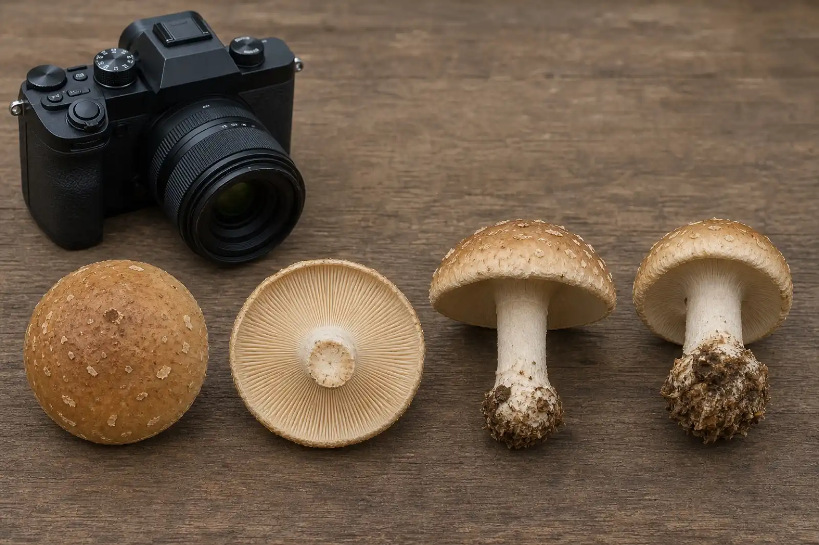
Mushroom Photography Checklist for Identification
Capture the cap, underside, complete stem base, habitat, scale, and specimens at different ages before asking for identification help.
identification
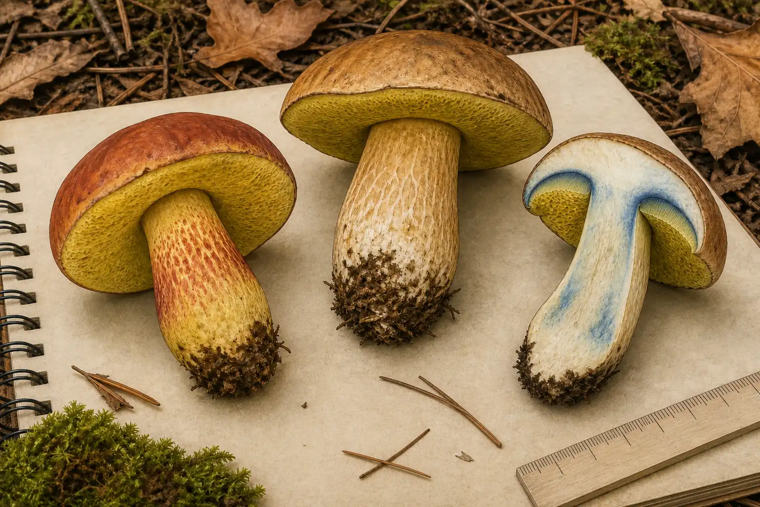
Bolete Identification: Pores, Staining, and Lookalikes
Learn how to document a bolete's pores, flesh staining, stem, habitat, and lookalikes without treating any one feature as an edibility test.
identification
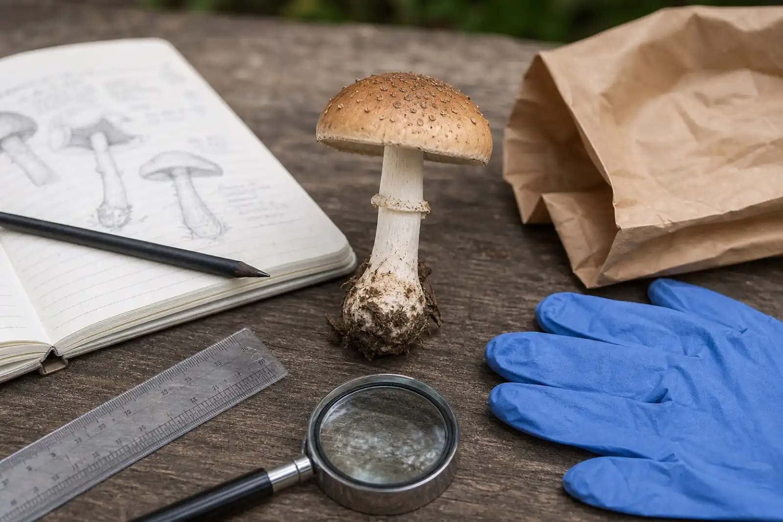
Mushroom Identification Safety Checklist
Use a conservative checklist before handling, documenting, or discussing any wild mushroom, especially when edibility is being considered.
Continue exploring
Browse more practical guides in Identification or visit the mushroom encyclopedia.