Identification
Edible Mushroom Lookalikes: A Safer Comparison Method
Compare edible mushroom lookalikes by complete structure, habitat, development, and regional evidence without treating a web page as eating clearance.
Safety first
Use as a field-record guide
Document the whole mushroom, habitat, substrate, and region before comparing names or photographs.
Open the identification safety hub →Decision limit
Do not use this as an edibility verdict
Wild mushroom consumption decisions require qualified local, in-person confirmation of the complete specimen.
Read the safety disclaimer →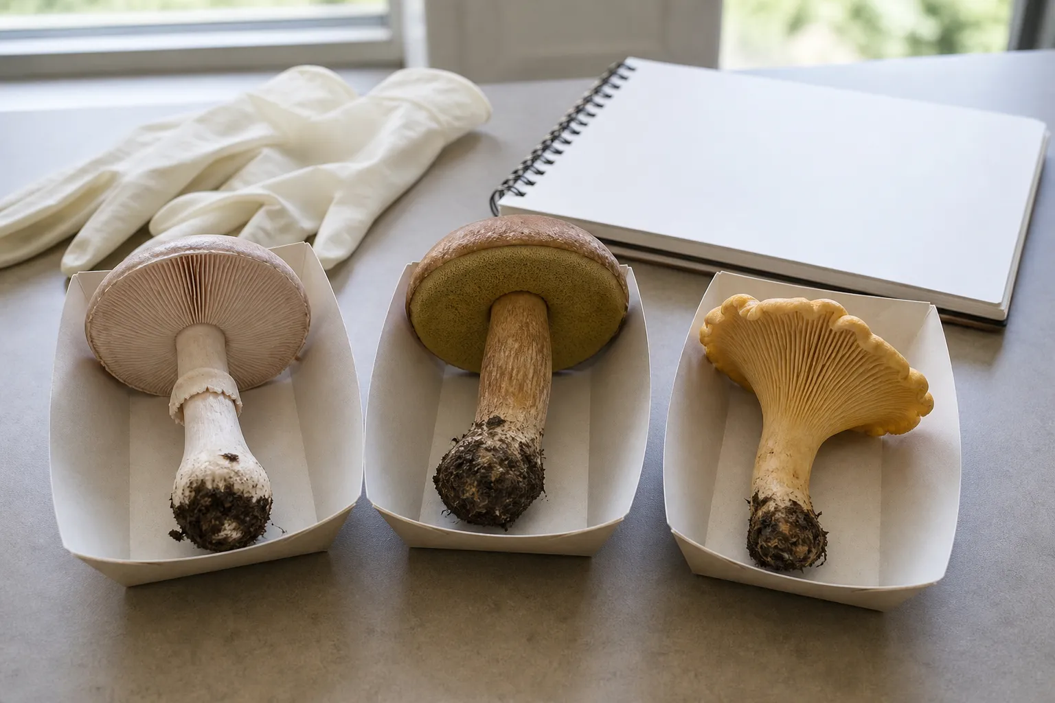
Safety scope: This editorial guide teaches comparison and documentation. It cannot confirm that a wild mushroom is edible, and it has no named expert or clinical reviewer. Consumption decisions require qualified local, specimen-based identification.
“Edible-looking” is not an identification category
There is no universal mark of edibility. Bright color does not reliably mean poisonous; dull color does not mean safe. Insects or mammals feeding on a mushroom do not predict human tolerance. A cap that peels, a pleasant smell, a silver spoon that stays bright, or thorough cooking cannot rule out dangerous toxins. These folk tests fail because unrelated fungi can share superficial traits while differing in chemistry.
A safer question is not “does this look edible?” but “what complete set of observations supports this taxon, and what dangerous alternatives remain?” The burden is asymmetric. Several matching characters may support a hypothesis, while one incompatible feature—such as a saclike volva at the base—can reject it. The method below organizes evidence; it does not provide eating clearance. Start with the broader mushroom identification workflow and use this page when an edible species name has already generated a comparison set.
Collect the evidence that photographs usually omit
Document more than the cap. Photograph the habitat before moving the specimen, then collect the entire fruit body, including the stem base and attached substrate. Record whether it grew from soil, buried wood, a standing trunk, a stump, dung, litter, or a living host. Note whether fruit bodies were solitary, scattered, clustered from one base, or arranged in arcs.
For gilled mushrooms, record gill attachment, spacing, thickness, branching, mature color, and bruising. Describe the stem surface, internal texture, ring, bulb, volva, rooting extension, and any veil remnants. For boletes, record pore color and size, pore-tube depth, cap texture, stem reticulation or dots, and staining separately on pores, cap flesh, and stem flesh. For brackets, inspect the fertile underside rather than relying on zoned cap color. For enclosed forms, cut vertically from top through base to look for a developing internal cap and stem.
Time is evidence. Young gills can be a different color from mature gills; latex and bruising may change over seconds or minutes; a ring may collapse; rain may wash veil patches away. Photograph the same feature immediately and again after a recorded interval. The mushroom photo checklist helps build a set that another person can evaluate.
Chanterelles versus orange true-gilled mushrooms
True chanterelles have blunt, decurrent folds or ridges rather than thin, blade-like gills, but that distinction needs a side-lit close view and a cut specimen. The folds often fork and cross-connect; the interior flesh is typically pale and solid. Growth from soil in association with trees fits their mycorrhizal ecology. Color alone is weak because weather, age, camera white balance, and several unrelated orange fungi overlap.
Jack-o’-lantern mushrooms (Omphalotus species) have true gills and commonly fruit in dense clusters from wood, including buried roots. False chanterelles and other orange-gilled species add regional complexity. Bioluminescence is not a practical field test: it may be faint, condition-dependent, and absent from an ordinary observation. A responsible comparison uses hymenium structure, substrate, grouping, internal flesh, geography, and a regional key together. If the underside is missing, blurred, or not mature enough to interpret, the evidence set is incomplete.
Morels versus chambered or folded lookalikes
A true morel hypothesis should be tested with a full longitudinal section. Cap attachment, the relationship between cap and stem, and the internal cavity are more informative than a “brainlike” silhouette. Species and regional groups vary, so a single slogan such as “hollow means safe” is not sufficient. Gyromitra and other lookalikes can differ in chambering, attachment, lobing, and internal cottony tissue, yet weathered specimens and photographs can obscure those distinctions.
Toxicity within false-morel groups is not resolved by a history of local consumption or by parboiling advice found online. Chemistry can vary by species, specimen, preparation, and region, and cooking fumes can matter for some toxins. Do not use this guide to authorize eating a morel-like collection. Preserve an intact specimen and cross-section for a local expert who knows the regional taxa.
Agaricus versus Amanita and other pale gilled fungi
Market buttons can make a white, gilled wild mushroom feel familiar, but the genus Agaricus contains both edible and problematic species, and pale mushrooms occur in dangerous groups. Mature Agaricus gills generally progress toward chocolate brown and its spore deposit is brown. An Amanita can retain white gills and a pale spore deposit, with universal-veil evidence at the stem base. Cutting the stem above ground can remove the most important distinction.
Within Agaricus, record odor at the cap center and stem base, the location and speed of yellow or reddish staining, ring architecture, mature gill color, habitat, and regional species. “Smells like store mushrooms” is not enough. Chemical or phenolic odors can be difficult to perceive, and staining can be weak in dry or old material. A disagreement among traits means the specimen stays unidentified. Never infer “no volva” from a photograph that does not show an excavated base; the correct observation is “base not documented.”
Puffballs versus immature gilled mushrooms
Every puffball candidate should be sliced from top through the point of attachment. A uniform, solid white interior is expected only at an appropriate immature stage; yellowing, browning, powdering, or soft breakdown indicates development or decay. More importantly, an outline of a cap, gills, or stem inside reveals an unopened mushroom rather than a puffball. Some dangerous Amanita begin as button-like structures enclosed by a universal veil.
External shape and whiteness cannot substitute for the cut. Photograph both halves in neutral light, include the base, and note whether the interior is truly homogeneous. Even after that check, genus-level differences, regional toxic species, age, contamination, and individual tolerance remain relevant. A cut is a rejection tool, not a universal edibility certificate.
Boletes require a regional matrix, not one staining rule
“Avoid every blue-staining bolete” discards many taxa but still does not prove the remaining mushrooms safe. Conversely, “blue staining is harmless” ignores dangerous regional species. Bolete comparison should combine cap and pore color, pore shape, tube depth, stem ornamentation, basal color, flesh color, staining location and timing, tree associates, and geography.
Record a timed stain sequence: immediately after handling, after about one minute, and later. Separate pore bruising from cut-flesh changes. Photograph the stem at several heights because reticulation may be confined to the apex while dots or color concentrate lower down. Consult a current local key; a rule developed for European species may fail in North America, Asia, or Australia.
Bracket fungi: the fertile surface carries the comparison
Concentric bands appear in turkey tail, false turkey tail, and many other thin brackets. Turn every specimen over. True turkey tail candidates should have a poroid fertile surface, while some close visual mimics have a smooth or toothed underside. Record pores per millimeter with a scale, cap hairiness, thickness, flexibility, attachment, wood type, and decay context. Age and algae can change the upper surface dramatically.
The dedicated turkey tail species guide provides a taxon-focused account. This comparison illustrates the general rule: a conspicuous cap pattern is often less diagnostic than a quiet underside feature.
Regional names and seasons can mislead
Common names may refer to different species across countries, while taxonomic revisions split an old familiar name into regional lineages. Imported field guides can omit the local dangerous comparison. Fruiting season is supporting context, not proof; rainfall, elevation, irrigation, and unusual weather shift timing.
Write down country, state or province, approximate elevation, habitat, host or nearby tree, and date. Use a current regional key and, when consumption is contemplated, show physical material to a qualified local identifier. An app result or online vote can suggest what to investigate, but neither establishes that every dangerous alternative was excluded.
Build a comparison table before choosing a name
Create rows for the proposed species and each plausible dangerous or confusing alternative. Use columns for cap, fertile surface, stem, base, veil, internal section, spore deposit, odor, timed staining, substrate, host, grouping, season, and region. Mark each cell observed, not observed, unknown, or conflicting. “Unknown” must not be silently converted into a match.
Choose the next observation by information value. If two candidates differ primarily at the stem base, collecting another cap photograph adds little. If a bracket pair differs underneath, a pore-scale image matters more than a scenic habitat view. If the candidates require microscopy or chemistry, acknowledge that limit instead of improvising a visual shortcut. The identification safety checklist provides a final stop/go framework.
What to do after suspected poisoning
Symptoms after wild-mushroom ingestion are a medical problem, not an identification contest. Contact emergency services or the relevant poison center promptly; in the United States, Poison Help is 1-800-222-1222. Do not wait for symptoms to become severe and do not induce vomiting unless a medical professional directs it.
When safe, retain uncooked specimens, trimmings, cooked leftovers, packaging, photographs, collection location, meal time, symptom timeline, and a list of everyone who ate the meal. Keep material separated and refrigerated as directed so an expert can examine it. Do not rely on a social-media identification before seeking care. Different diners can have different onset and severity, and delayed syndromes can be especially serious.
Frequently asked questions
Is there a universal test for an edible mushroom?
No. Color, animal feeding, cap peeling, silverware reactions, smell, and cooking are not universal safety tests.
Why must the stem base be collected?
A buried bulb, saclike volva, rooting extension, or attached substrate can separate dangerous groups. Cutting the stem at ground level destroys that evidence.
Can photographs rule out a poisonous lookalike?
Usually not alone. Complete material, developmental stages, spore deposit, microscopy, chemistry, host evidence, and regional expertise may be required.
Are false gills enough to identify a chanterelle?
No. Blunt folds are useful, but growth pattern, flesh, odor, substrate, geography, and exclusion of true-gilled lookalikes must agree.
What should be saved after suspected mushroom poisoning?
Seek urgent professional help and, when safe, preserve specimens, leftovers, photographs, packaging, location, and timing.
References
- U.S. Health Resources and Services Administration. Poison Help. Immediate U.S. poison-center access and emergency guidance.
- North American Mycological Association. Mushroom Poisoning Syndromes. Syndrome-oriented toxicology and response context.
- Centers for Disease Control and Prevention. Wild mushroom poisoning outbreak report. Evidence that informal identification can lead to severe poisoning.
- Queensland Poisons Information Centre. Mushroom poisoning. Public-health advice emphasizing prompt professional assessment.
Source quality notes
MushroomScope cites sources that match the page scope, such as taxonomic databases, extension guidance, food-safety agencies, food-composition databases, and peer-reviewed or institutional health references. Sources support context and uncertainty; they do not turn an online page into specimen identification, medical advice, or a tested recipe record.
Frequently asked questions
Is there a universal test for an edible mushroom?
No. Color, animal feeding, cap peeling, silverware reactions, smell, and cooking are not universal edibility tests.
Why must the stem base be collected?
A buried bulb, saclike volva, rooting extension, or attached substrate can separate dangerous groups, and cutting the stem at ground level destroys that evidence.
Can photographs rule out a poisonous lookalike?
Usually not by themselves. Complete material, developmental stages, spore deposit, microscopy, chemistry, host evidence, and regional expertise may be required.
Are false gills enough to identify a chanterelle?
No. Blunt folds are useful, but growth pattern, flesh, odor, substrate, geography, and exclusion of true-gilled lookalikes must agree.
What should be saved after suspected mushroom poisoning?
Seek urgent professional help and, when safe, preserve uncooked specimens, cooked leftovers, photographs, packaging, collection location, and meal timing.
Related guides
identification
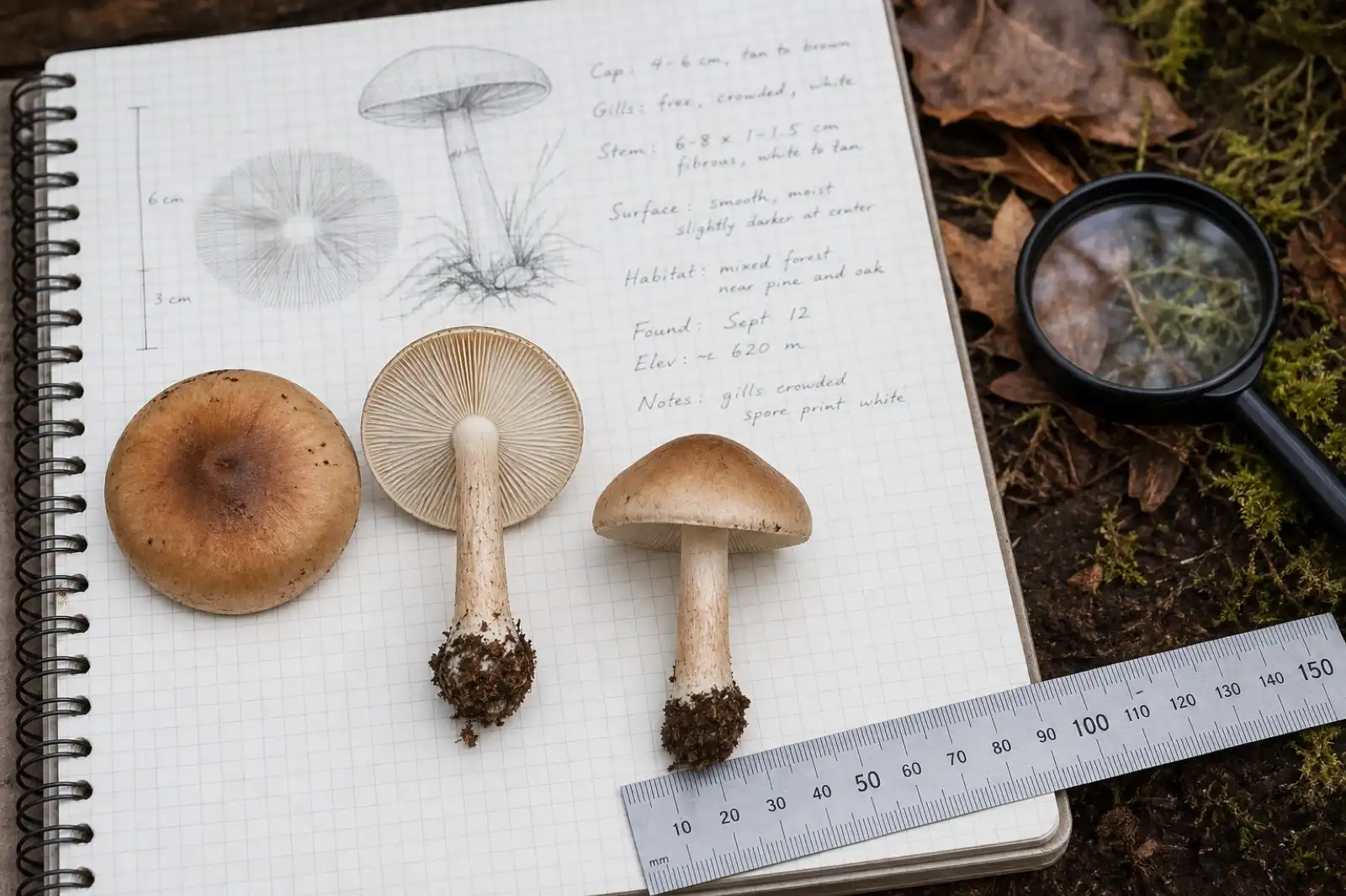
How to Identify Mushrooms: A Beginner's Field Guide
Build an identification from the whole specimen, substrate, habitat, spore deposit, and location—not from a single photograph.
identification
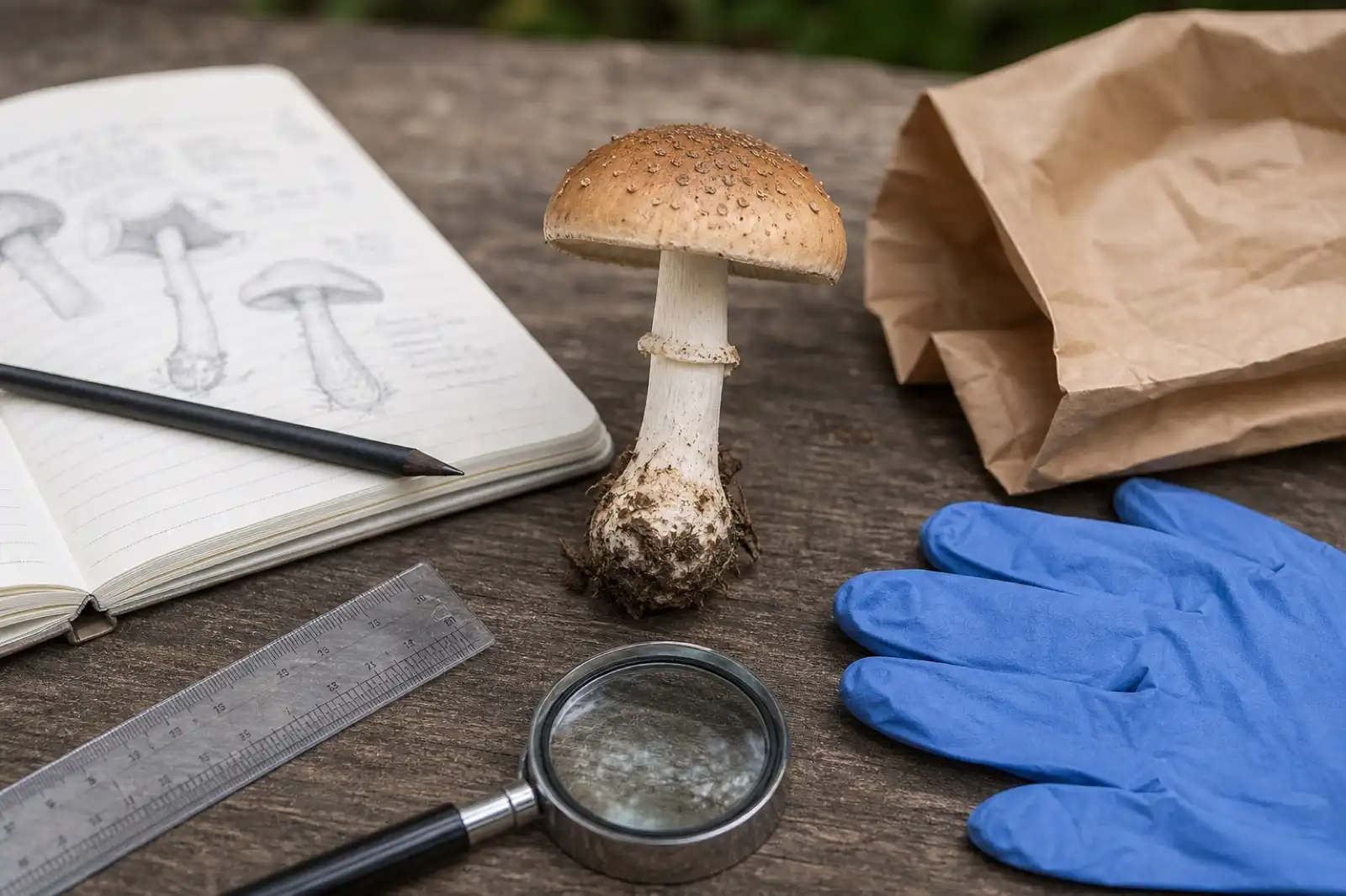
Mushroom Identification Safety Checklist
Use a conservative checklist before handling, documenting, or discussing any wild mushroom, especially when edibility is being considered.
identification
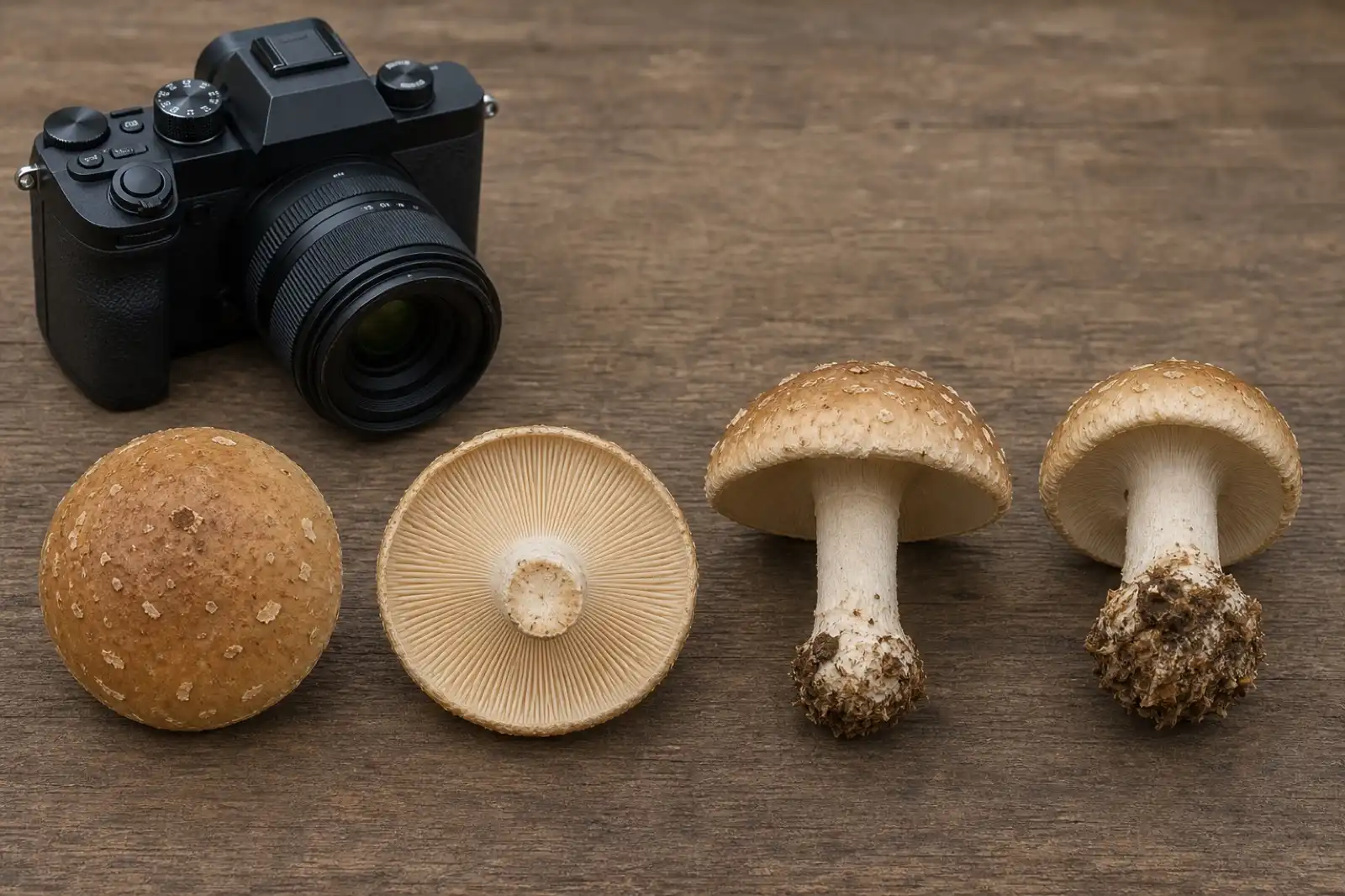
Mushroom Photography Checklist for Identification
Capture the cap, underside, complete stem base, habitat, scale, and specimens at different ages before asking for identification help.
Continue exploring
Browse more practical guides in Identification or visit the mushroom encyclopedia.