Mushroom Encyclopedia
Witch's Butter Fungus: Yellow Jelly, Hosts, and Lookalikes
Document witch's butter by yellow gelatinous folds, wet-to-dry change, hardwood attachment, host-fungus ecology, microscopy, and jelly-fungus lookalikes.
By MushroomScope Editorial Team · · Updated
Species context
Treat profiles as reference material
Species profiles summarize documented traits, taxonomy, and lookalikes, but natural variation can be substantial.
Use the safety checklist →Decision limit
A profile cannot authenticate a specimen
Do not eat a wild mushroom from an online match, single feature, or generated image.
Read the safety disclaimer →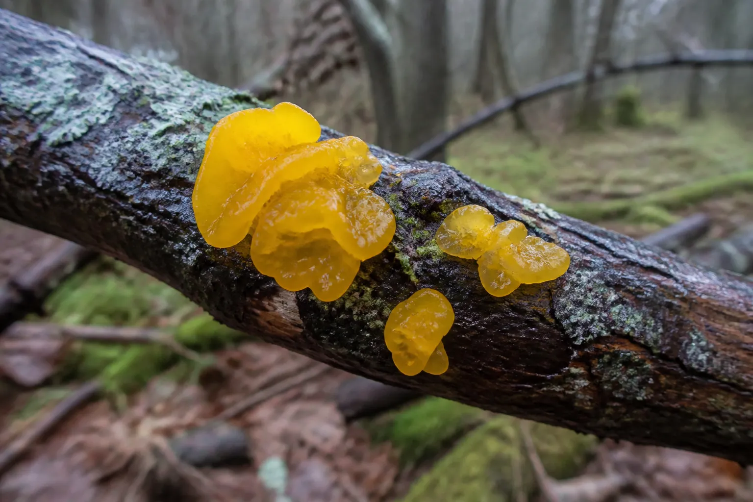
Quick facts
- Scientific name
- Tremella mesenterica
- Common names
- Witch's butter, Yellow brain fungus, Golden jelly fungus
- Taxonomy
- Tremella · Tremellaceae
- Edibility context
- Unknown: No food recommendation is made; a yellow jelly form does not establish identity, substrate cleanliness, or individual safety.
- Spore print
- White to pale, usually less practical than microscopy of basidia and spores for identification.
- Toxicity context
- Unknown: Do not infer safety from folklore, small size, or the lack of a famous poisoning syndrome.
- Season
- January, March, October, December
- Growing difficulty
- Expert
- Regions
- widespread temperate records, local occurrence requires verified regional data
Safety limit: These labels summarize documented context; they never authenticate a wild specimen. Cooking does not make a poisonous species safe.
Witch’s butter can change from a nearly invisible orange crust in dry weather to a bright folded jelly after rain. That transformation explains many “appeared overnight” reports. The fruit body was often already present, shrunken on the branch, then rehydrated when water returned.
The deeper ecological story is equally important: Tremella mesenterica is not simply feeding on wood. It parasitizes the mycelium of wood-decaying crust fungi, commonly species of Peniophora. Identification therefore connects a yellow jelly to hardwood, weather, a possible fungal host, and microscopic evidence.
Observe the fungus before hydration changes
Photograph the branch and fruit body under current conditions. Note recent rain, frost, sun exposure, and whether lobes are fully expanded. If the site can be revisited without disturbance, repeat the same frame after a dry interval.
Do not pour water onto an unknown fungus merely to stage a transformation. Natural time-series observations preserve ecology and avoid moving spores or contaminants.
Describe lobes rather than calling them a brain
Hydrated fruit bodies form overlapping gelatinous folds, leaflike flaps, or irregular cushions. The common phrase “yellow brain” is visually convenient but imprecise. Record total width, lobe thickness, translucency, branching, and whether separate clusters merge.
Fresh color ranges from lemon yellow through golden orange. Camera processing can turn a restrained yellow into neon. Include a neutral reference and use diffuse light.
Confirm direct emergence from bark
The jelly typically appears through cracks in dead hardwood bark. Photograph whether the branch is fallen, attached but dead, or part of a stump. Identify hardwood only from independent tree characters, not from the fungus’s expected ecology.
Look closely for a thin crust fungus on or beneath nearby bark. A Peniophora host can be inconspicuous, and absence from a photograph is not evidence that it is absent biologically.
Treat the host fungus as part of the record
The parasitic interaction distinguishes the ecology from an ordinary saprotrophic jelly. Where permitted, a careful sample can include the jelly, its exact bark attachment, and adjacent crust tissue. Photograph layers before separating anything.
Naming the crust fungus may require its own microscopy. Do not label any nearby paint-like patch Peniophora solely because witch’s butter is expected to parasitize that genus.
Map the interaction across the branch
A close photograph of yellow lobes loses the spatial evidence that makes this fungus ecologically distinctive. Build a branch map before sampling: mark the jelly clusters, visible crust margins, bark cracks, exposed wood, branch ends, and areas without fruit bodies. Photograph the entire branch, then overlapping close frames that preserve those relationships.
The map supports three different claims that should not be collapsed. Direct emergence through bark describes where the jelly fruit body appears. A neighboring crust suggests a candidate host but does not establish its identity. Microscopic or culture evidence can support an interaction only when the samples remain traceable to their exact positions. Merely finding Peniophora somewhere on the same fallen limb is not proof that the photographed jelly parasitized that colony.
When a qualified project samples the interaction, assign separate identifiers to jelly tissue, adjacent crust, bark interface, and controls. Photograph each removal point and record distance between samples. Mixed DNA from an interface can contain the jelly fungus, its host, endophytes, yeasts, and environmental contaminants; a sequence list is not automatically a map of biological roles.
Repeated non-destructive visits add evidence unavailable from one collection. The same framed view after rain and after drying can show whether particular lobes re-expand and whether the visible crust boundary changes. It cannot, by itself, prove growth rate or parasitism, but it prevents hydration-driven volume from being reported as new biomass.
Track the dry state
As water leaves, lobes collapse and color deepens. A cluster can become a thin tough film that looks unrelated to the hydrated stage. Rain may expand it again, although repeated weathering eventually damages the tissue.
Record dimensions wet and dry rather than claiming that a specimen grew several centimeters in a day. Water-driven volume change is not the same as new biomass production.
Use microscopy for yellow jelly comparisons
Tremellales characteristically have basidia divided by longitudinal septa, sometimes described as cruciate. Spores and conidiogenous stages can also contribute evidence. Dacrymyces has a different basidial architecture and frequently occurs on conifer wood.
These characters require correctly prepared mature tissue and competent interpretation. Preserve hydration history and substrate with a sample; a detached yellow fragment loses valuable context.
Compare Dacrymyces with substrate and microstructure
Large orange-yellow Dacrymyces cushions can resemble witch’s butter. Conifer attachment should redirect attention, while hardwood supports but does not prove Tremella. Fruit-body shape overlaps too much for color sorting.
Microscopic basidia provide a stronger division than “more orange” or “more folded.” If microscopy is unavailable, use a broader jelly-fungus record rather than an invented certainty.
Compare other Tremella-like yellow fungi
Names such as Tremella aurantia and yellow cultivated jelly fungi appear in guides and markets. Their fungal hosts, geographic distributions, and modern generic placements differ. A product label also represents a supply-chain claim, not a field identification method.
Keep exact scientific names attached to exact sources. Do not transfer medicinal, culinary, or cultivation statements across yellow jelly taxa merely because photographs look alike.
Distinguish witch’s butter from wood ear
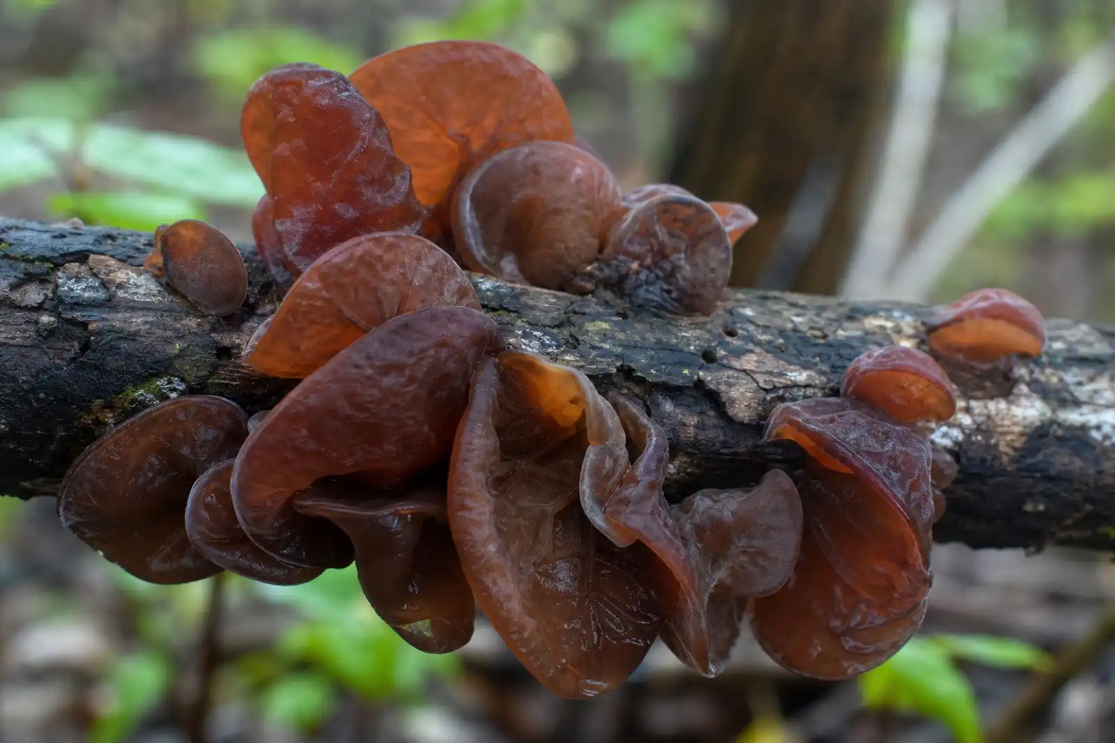
Wood ear and cloud ear fungi are brown to dark, ear- or cup-shaped gelatinous fruit bodies on wood. Their fertile surfaces, thickness, texture, and taxonomic placement differ. The wood ear guide addresses the market-name and species boundary separately.
The shared gelatinous texture is an adaptation and field clue, not evidence that all jelly fungi are interchangeable ingredients.
Distinguish it from jelly tooth fungi
Jelly tooth fungi carry distinct downward teeth on a translucent fruit body, often on conifer wood. Witch’s butter has folded lobes without a toothed underside. This comparison shows why a close underside photograph matters even in soft gelatinous fungi: a translucent top view can conceal an entire field of projections.
Check for teeth before handling; backlighting can make small projections clearer.
Record weather more carefully than season
Witch’s butter may be noticed in winter, spring, or autumn depending on regional moisture. A month list is less explanatory than rain, thaw, humidity, and branch exposure. Old dried tissue can persist through periods when it is not actively expanding.
Record air temperature, precipitation over the previous days, and whether the branch is shaded or exposed. Those details support repeat observations without making climate claims from one patch.
Build a host-aware observation workflow
- Photograph the whole branch and canopy context.
- Determine hardwood versus conifer with independent evidence.
- Measure hydrated clusters and individual lobes.
- Record translucency, color, elasticity, and dry-state remains.
- Photograph adjacent crust fungi and the exact emergence point.
- Preserve a legal contextual sample for microscopy if needed.
- Compare basidia, spores, substrate, and host fungus with a regional key.
The habitat guide helps prevent a fungus-on-fungus interaction from being reduced to “yellow growth on a stick.”
Do not convert folklore into evidence
The common name has accumulated stories about witches, weather, food, and remedies. Historical naming can be culturally interesting but does not verify taxonomy, dose, efficacy, or safety. A modern product claim needs product-specific identity and evidence.
This species guide does not diagnose disease or recommend topical, internal, or veterinary use. Gelatinous appearance is not proof of soothing or moisturizing effects.
Keep ingestion behind a firm boundary
Some informal references describe witch’s butter as edible but bland. This page makes no food recommendation. Close jelly fungi, substrate contaminants, polluted wood, microbial spoilage, and individual reactions are not resolved by a yellow photograph.
Never sample a field jelly to test texture. Cooking cannot repair misidentification or remove every environmental contaminant. If illness follows wild fungus consumption, contact local emergency services or a poison center and retain specimens and timing details.
Explain why cultivation is unusually complex
Because T. mesenterica depends on another fungus, a verified project must maintain at least two authenticated biological actors and demonstrate their interaction. A yellow blob appearing in a culture is not proof of successful fruiting; bacteria, yeasts, slime molds, and other fungi can create wet colored growth.
Research records should include both culture accessions, substrates, inoculation sequence, controls, temperature, water activity, microscopy, and contamination exclusions. That is an expert co-culture problem, not a generic hardwood bag recipe.
Observe without stripping the branch
Small jelly fungi can be documented in place through several wet-dry cycles. Take minimal material only where legal and scientifically useful. Leave the host crust and some fruit bodies intact.
Avoid breaking attached dead limbs overhead. A fungal observation never justifies unsafe access to a branch or climbing a compromised tree.
Preserve wet tissue for the question being asked
Gelatinous fungi are easily flattened into an uninformative film. Before preservation, allocate material deliberately: photograph a fresh cross-section, place a small portion in the medium required by the receiving specialist, and dry a separate contextual voucher with branch and host-crust notes. Do not assume that one storage method serves microscopy, culture, and DNA equally well.
Label wet and dry measurements separately and record the interval since rain. If a culture is attempted, maintain sterile technique and retain an uninoculated control; growth from a field fragment may represent the crust host, an environmental yeast, or a contaminant rather than the visible jelly. A culture result without re-identification should never be used to promote a household cultivation method.
Evidence and image limits
GBIF and NCBI provide taxonomic and occurrence checkpoints. The GBIF Peniophora record frames the host-fungus group but does not prove the host of an individual collection. NAMA supports conservative emergency communication.
The cover is an original AI editorial image. It was checked for translucent yellow folded lobes, direct hardwood emergence, wet bark, natural light, realistic scale, clarity, and absence of text or watermark. It is not proof of host fungus, microscopy, or species. No named mycologist or clinician reviewed this page; its review status is editorial.
Frequently asked field questions
Can the fungus survive drying?
Fruit bodies can rehydrate after drying, but repeated extremes cause deterioration. Re-expansion does not mean indefinite viability or fresh condition.
Is brighter yellow a younger specimen?
Not reliably. Hydration, light, weathering, camera balance, and taxon all affect color. Use structural and ecological evidence.
Does it only grow on fallen branches?
No. It may appear on dead portions of standing or attached hardwood as well as fallen debris, wherever the fungal host occurs.
Can the associated crust be seen without a microscope?
Sometimes a thin crust is visible, but species identification commonly needs microscopic work. A bare-looking branch may still contain host mycelium.
Why not list medicinal benefits?
Evidence about a compound, a different yellow jelly species, or a commercial extract cannot be transferred to a field collection of unresolved identity.
References
- GBIF — Tremella mesenterica
- NCBI Taxonomy Browser — Tremella mesenterica
- GBIF — Peniophora
- North American Mycological Association — Mushroom Poisoning Syndromes
Editorial review: September 5, 2026. Taxonomy, host-fungus boundaries, source URLs, identification limits, and poison-response wording were checked. Corrections can be sent through the site contact page.
Source quality notes
MushroomScope cites sources that match the page scope, such as taxonomic databases, extension guidance, food-safety agencies, food-composition databases, and peer-reviewed or institutional health references. Sources support context and uncertainty; they do not turn an online page into specimen identification, medical advice, or a tested recipe record.
Frequently asked questions
Why does witch's butter appear after rain?
Its gelatinous tissue rehydrates and expands in wet weather, becoming conspicuous; in dry periods the same fruit body may shrink into a thin dark orange film.
Does Tremella mesenterica decay wood by itself?
It is primarily parasitic on wood-decaying Peniophora fungi, so the branch supports a multi-organism interaction rather than a simple direct wood-decayer story.
How can witch's butter be separated from Dacrymyces?
Host wood, fruit-body form, microscopic basidia and spores, and the presence of an associated crust fungus matter; yellow color alone cannot separate jelly fungi.
Can a dried yellow smear rehydrate into witch's butter?
Some gelatinous fruit bodies shrink markedly and expand again with moisture, but a rehydration response is not unique enough to establish the species.
Is witch's butter edible?
This guide makes no food recommendation. Identity, substrate contamination, freshness, close species, and personal reactions cannot be resolved from a web photograph.
Related guides
mushrooms
Auricularia heimuer: Wood Ear Mushroom Species Guide
A practical guide to Auricularia heimuer identification, wood-decay ecology, log and bag cultivation, nutrition, rehydration safety, and FAQs.
mushrooms
Birch Polypore: Fomitopsis betulina Host and Decay Guide
Identify birch polypore through verified birch host evidence, annual bracket development, fine white pores, brown-rot context, and perennial lookalike separation.
identification
Using Habitat to Identify Mushrooms
Record substrate, associated plants, soil, wood type, moisture, elevation, and fruiting pattern as identification evidence.
Continue exploring
Browse more practical guides in Mushroom Encyclopedia or visit the mushroom encyclopedia.