Mushroom Encyclopedia
Scarlet Elf Cup: Red Cups, Dead Wood, and Species Limits
Document scarlet elf cups by red cup shape, pale outer surface, buried hardwood attachment, spring timing, microscopy limits, and regional Sarcoscypha species.
By MushroomScope Editorial Team · · Updated
Species context
Treat profiles as reference material
Species profiles summarize documented traits, taxonomy, and lookalikes, but natural variation can be substantial.
Use the safety checklist →Decision limit
A profile cannot authenticate a specimen
Do not eat a wild mushroom from an online match, single feature, or generated image.
Read the safety disclaimer →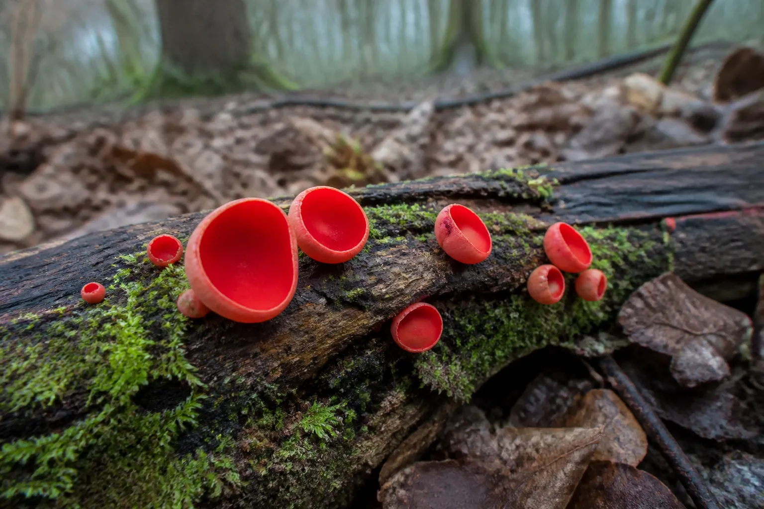
Quick facts
- Scientific name
- Sarcoscypha coccinea
- Common names
- Scarlet elf cup, Scarlet cup, Scarlet cup fungus
- Taxonomy
- Sarcoscypha · Sarcoscyphaceae
- Edibility context
- Unknown: No culinary recommendation is made; close-species resolution and specimen condition are not established by a web image.
- Spore print
- Usually not collected as a conventional cap print; microscopic ascospore examination uses discharged spores or prepared tissue.
- Toxicity context
- Unknown: Absence of a famous toxin is not evidence that an unidentified red cup, contaminated substrate, or individual serving is safe.
- Season
- January, February, March, April
- Growing difficulty
- Expert
- Regions
- Europe, temperate occurrence regions represented by verified records
Safety limit: These labels summarize documented context; they never authenticate a wild specimen. Cooking does not make a poisonous species safe.
Scarlet elf cups appear at a season when woodland color is scarce, making a red cup on a mossy branch feel instantly recognizable. The color and shape identify a useful comparison group, but they do not reliably distinguish Sarcoscypha coccinea from close relatives such as S. austriaca. A responsible page must therefore explain both the visible pattern and the point at which microscopy becomes necessary.
This guide uses S. coccinea as the focal name while keeping the genus-level boundary explicit. It does not approve a food use or claim that a cover image can resolve the species.
Confirm that the cup grows from wood
Start with the substrate. Follow the base through moss and leaves until it reaches a decaying hardwood twig or branch. Work gently; an attachment can be long and partly buried. Photograph the wood in place, its diameter, decay stage, bark remnants, and surrounding tree community.
A cup that seems to grow from soil may still originate from buried wood. Conversely, nearby wood is not proof of attachment. The habitat guide provides a practical method for distinguishing visible substrate from an assumed host.
Read the inner and outer surfaces separately
The vivid scarlet inner face is smooth because it is a spore-bearing hymenium, not a gilled cap. The outside is paler—often whitish, pinkish, or light orange—and may look finely hairy under magnification. Record both surfaces in neutral light because phone processing can exaggerate red saturation.
Use a scale and avoid crushing the rim. Photograph from above, from the side, and at the attachment. A single overhead red circle hides the very characters needed for comparison.
Follow cup development instead of expecting one shape
Young specimens can be deep goblets with an inward-curved rim. As they expand, the cups become shallow, irregular, split, or almost saucer-like. Rain, frost, drying, neighboring fruit bodies, and the angle of the supporting twig distort symmetry.
Measure diameter and depth across several stages. “Perfect cup” is a photographic preference, not a taxonomic requirement. A series also reveals whether small discs are juveniles or a separate species fruiting beside larger cups.
Understand how an ascomycete releases spores
Scarlet cups belong to the Ascomycota. Spores develop inside microscopic sacs called asci arranged in the inner hymenium. Under favorable conditions they may be discharged into the air. This differs from a conventional gilled mushroom dropping spores from basidia beneath a cap.
A standard overnight cap print is therefore not the most informative method. Microscopic work may examine discharged mature spores, ascus features, and tissue from the outer surface. Collect only where permitted and with a plan for preservation and examination.
Separate S. coccinea from S. austriaca honestly
These two names are frequently confused because cup color, size, substrate, and season overlap. Regional keys may use mature spore dimensions and shape, oil droplets, germination behavior, and whether outer hairs are straight or conspicuously coiled. Those characters are not visible in an ordinary habitat photograph.
If microscopy is unavailable, “Sarcoscypha species” can be the most accurate result. Do not use geography alone as a substitute; occurrence ranges overlap and records can be incomplete or historically misidentified.
Keep additional Sarcoscypha names regional
North American comparison sets can include S. occidentalis and other taxa. A smaller, more stalked cup may suggest a different route through a local key, but size and stalk visibility change with age and burial. Imported names from a European photo gallery may not fit the local species complex.
Record country, region, elevation, habitat, and the literature used. Scientific value comes from a transparent level of confidence, not from forcing every observation to a species.
Compare orange peel fungus by ecology and structure
Aleuria aurantia often forms orange irregular cups or flattened patches on disturbed soil, path edges, and bare ground. Its color can approach red in photographs, while faded Sarcoscypha can appear orange. Substrate and the contrast between inner and outer surfaces help redirect the comparison.
Other red or orange discs grow on burned soil, dung, plant stems, and wood. A two-species image comparison is never exhaustive. Use a current regional cup-fungus key.
Use season as context, not a shortcut
Winter through early spring is characteristic in many temperate observations. Snowmelt, rainfall, prolonged cool humidity, and decay moisture can make the fruit bodies conspicuous before leaf-out. Maritime sites, high elevations, and different latitudes shift the period.
Record recent weather and whether cups are fresh, frost-damaged, or bleached. An old specimen can remain visible after active spore release has ended.
Document the surrounding microhabitat
Photograph canopy and shrub layers, slope, drainage, litter depth, moss, and the position of wood relative to soil. Note whether a branch lies across a wet hollow, stream margin, upland floor, or managed path. These details can explain moisture without claiming that one microhabitat proves a name.
Avoid peeling bark or turning every branch in a productive patch. Cup fungi are small, and repeated disturbance can destroy both material and context.
Record the wood as carefully as the cup
“Hardwood” is a starting category, not a complete substrate record. Photograph bark that remains, end grain, twig branching, buds or attached leaves when present, and the point where the cup emerges. Record whether the wood is suspended, soil-contacting, waterlogged, or buried; also note diameter and a simple decay class such as firm, softened, or easily fragmented. Do not name a host tree from color alone.
The distinction matters because host associations reported in keys are only useful when the wood identification is credible. A branch lying beneath willow does not automatically belong to willow, and floodwater or garden maintenance can move debris. When the host cannot be resolved, retain “decaying hardwood branch” and preserve photographs that a botanist can revisit.
Link every microscopic preparation to that same collection and wood record. If two red-cup patches occupy different branches, number them separately even when they are only centimeters apart. This prevents a spore measurement from one collection being attached to habitat notes from another and makes a later revision from S. coccinea to a close species auditable.
Build a microscopy-ready record
- Photograph the group undisturbed with habitat context.
- Trace cups to their actual substrate.
- Record cup width, depth, color, outer texture, and stalk visibility.
- Include young and mature stages.
- Preserve a small legal sample promptly for microscopy.
- Note whether spores appear mature and how the sample was stored.
- Compare mature spores and outer hairs with a current regional key.
The mushroom photo checklist helps ensure that a beautiful overhead frame is accompanied by useful side and substrate views.
Avoid food claims from a decorative appearance
Scarlet cups are sometimes described in casual sources as edible or used as decoration. This draft makes no culinary recommendation. Tiny portions, uncertain identity, polluted roadside or treated-wood substrate, microbial condition, and individual reaction all complicate a simplistic label.
Do not taste an unknown cup. Never transfer a culinary statement about one Sarcoscypha name to an unresolved regional collection. Cooking does not repair a mistaken identity or remove every environmental contaminant.
Respond to exposure without guessing the name
If someone becomes ill after consuming a wild fungus, contact local emergency services or a poison center promptly. Preserve uncooked specimens, photographs, substrate notes, meal timing, and preparation details. A responder needs the evidence trail more than a confident common name.
NAMA’s syndrome overview is useful background, but symptoms overlap and online matching cannot replace medical evaluation.
Cultivation belongs to research, not a food shortcut
An ascomycete can sometimes be isolated in culture, but culture growth is not the same as verified fruiting. Substrate species, decay stage, temperature cycles, moisture, light, competitors, and strain identity may all influence cup production. A visually red contaminant cannot be accepted as the intended species.
Any study should retain culture provenance, microscopy, controls, and containment. This is not a beginner mushroom-growing project and should not be promoted as a home food method.
Preserve a population with minimal collection
Take only the few cups required for a defensible record. Leave immature and mature fruit bodies and avoid stripping an entire branch. Record precise locality privately where conservation or site sensitivity matters.
Repeated photography from the same marked angle can document expansion, frost damage, and senescence without collecting each stage. That time series may be more useful than a basket of detached cups.
Store a voucher without losing its context
If a qualified recorder requests material, assign a collection number before drying and keep field photographs, coordinates, substrate notes, measurements, and provisional name under the same identifier. Dry the small sample promptly with gentle airflow rather than sealing a wet cup in plastic, where bacteria and molds can destroy microscopic characters. Keep the wood fragment only when permission and curation needs justify it.
A useful voucher is not merely a dried red shape. The label should distinguish collector, identifier, collection date, identification date, microscope method, and the key or publication used. If a later specialist revises the name, retain the earlier determination in the record instead of erasing the history. That audit trail matters in a genus where old photographs and occurrence points may combine several close species.
Evidence and image boundaries
GBIF and NCBI provide name, occurrence, and lineage checkpoints; they do not certify a photographed specimen. Close-species statements are framed as microscopy problems rather than promises that a single macroscopic trick will work everywhere.
The cover is an original AI editorial image checked for smooth red inner cups, pale exteriors, woody attachment, spring woodland context, sharpness, natural color, and no visible text or watermark. It is not microscopy or voucher evidence. No named mycologist or clinician reviewed this draft.
Frequently asked field questions
Can frost change the cup color?
Yes. Freezing, drying, age, and camera processing can darken or bleach tissue. Record fresh color in diffuse light and note weather damage.
Are the tiny cups always young specimens?
No. They may be juveniles, a naturally small taxon, or a second species. Follow a marked group over time and compare attachment and microscopy.
Does a visible stalk rule out S. coccinea?
No. A short narrowed attachment may become visible when litter is removed. Stalk length alone is not enough to resolve close species.
Why photograph the outer surface?
Outer texture and hairs can guide microscopic sampling, and the pale exterior is part of the field profile. An inner-surface photo hides both.
Can an app distinguish the two common European species?
An app may suggest the genus, but it cannot measure mature spores or reliably evaluate outer-hair form from a general photograph.
References
- GBIF Secretariat. Sarcoscypha coccinea, species 8156721. Taxonomic and occurrence-record checkpoint; a database record does not identify the photographed cups.
- National Center for Biotechnology Information. Taxonomy Browser: Sarcoscypha coccinea. Lineage and linked sequence-record context; specialist literature remains necessary for nomenclature.
- GBIF Secretariat. Sarcoscypha austriaca, species 8689016. Separate close-species record used to keep the S. coccinea comparison explicit.
- North American Mycological Association. Mushroom Poisoning Syndromes. General exposure-response context; symptoms require medical or poison-center assessment rather than online species matching.
Last editorial review: September 2, 2026. This page received editorial review only; no named mycologist or clinician reviewed it. Recheck current regional keys, taxonomic records, and poison-response contacts before relying on the record.
Source quality notes
MushroomScope cites sources that match the page scope, such as taxonomic databases, extension guidance, food-safety agencies, food-composition databases, and peer-reviewed or institutional health references. Sources support context and uncertainty; they do not turn an online page into specimen identification, medical advice, or a tested recipe record.
Frequently asked questions
Is every red cup fungus Sarcoscypha coccinea?
No. Several Sarcoscypha species and unrelated cup fungi overlap in color and form; hairs, spores, host, region, season, and sometimes DNA separate them.
Where does a scarlet elf cup attach?
The cup typically arises from decaying hardwood twigs or branches, sometimes buried under litter so deeply that it appears to fruit directly from soil.
Why is the inside red but the outside pale?
The bright inner surface is the fertile hymenium, while the outer surface is usually paler and may carry hairs whose microscopic form can aid species comparison.
Can scarlet elf cups be identified from cap shape alone?
No. Cup shape changes with age and weather, and close species may be nearly indistinguishable without microscopic examination of spores and outer hairs.
Are scarlet elf cups edible?
This guide makes no food recommendation. Small cup fungi can be confused, polluted by their substrate, or poorly documented, and a photograph cannot establish safety.
Related guides
mushrooms
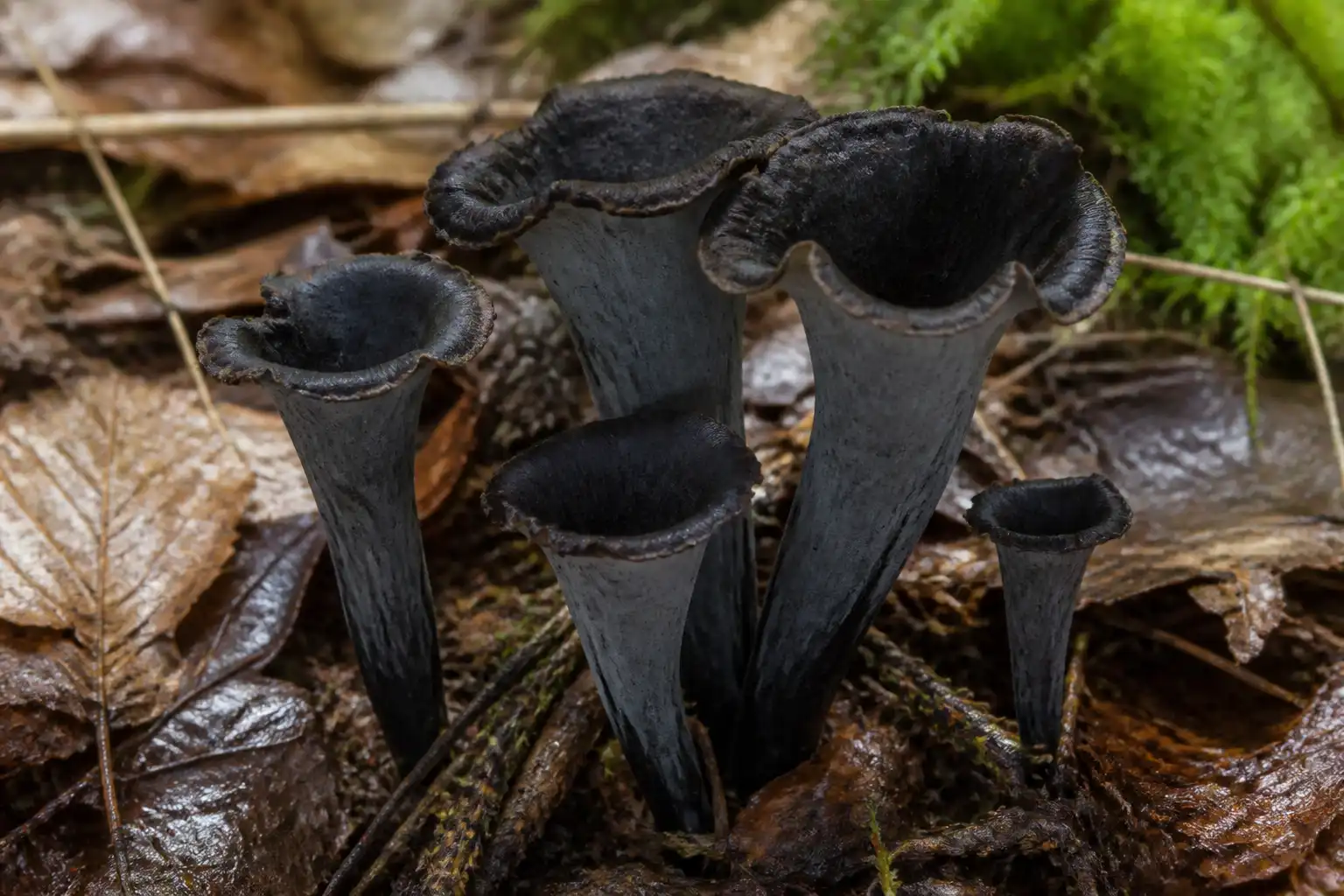
Craterellus cornucopioides: Black trumpet Species Guide
Learn Black trumpet identification, habitat, season, lookalikes, safety, food context, and cultivation limits with current taxonomy and sources.
identification
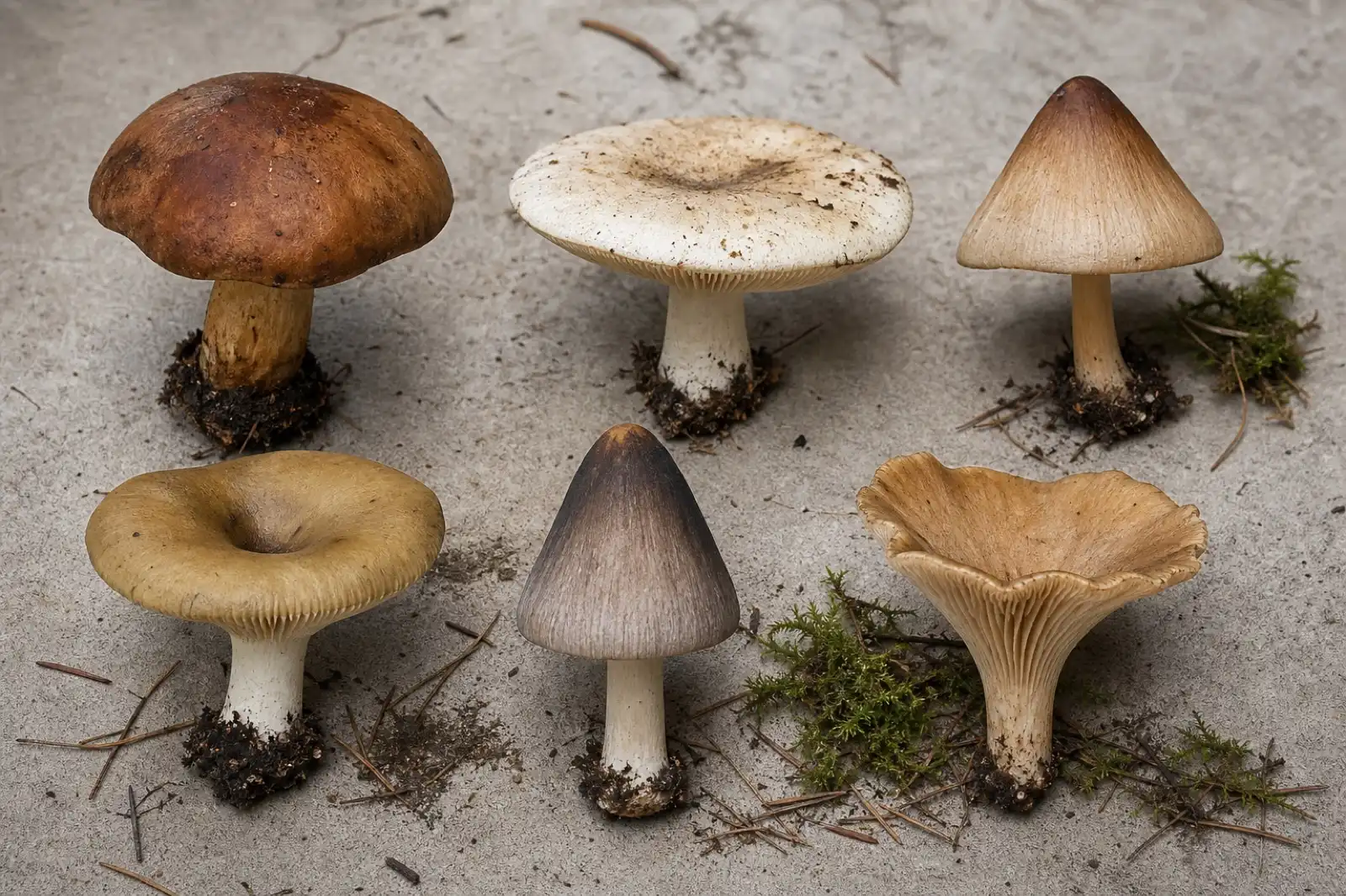
Mushroom Cap Shapes: A Practical Identification Guide
Compare mushroom cap shapes from convex and bell-shaped to flat, depressed, funnel-shaped, and uplifted, while recording age, margin, surface, and profile.
identification
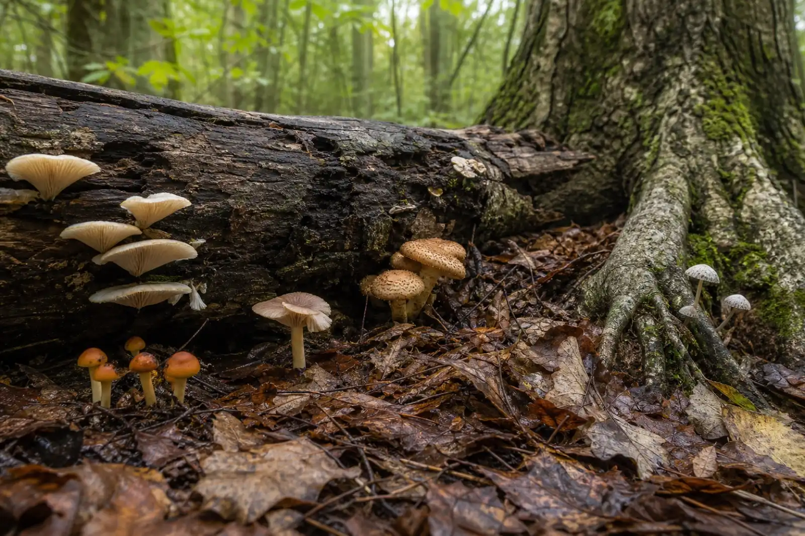
Using Habitat to Identify Mushrooms
Record substrate, associated plants, soil, wood type, moisture, elevation, and fruiting pattern as identification evidence.
Continue exploring
Browse more practical guides in Mushroom Encyclopedia or visit the mushroom encyclopedia.