Mushroom Encyclopedia
Honey Fungus: Armillaria mellea Identification and Tree Ecology
Identify honey fungus through clusters, rings, white spores, rhizomorphs, host evidence, and regional Armillaria limits, with conservative food and tree-risk guidance.
By MushroomScope Editorial Team · · Updated
Species context
Treat profiles as reference material
Species profiles summarize documented traits, taxonomy, and lookalikes, but natural variation can be substantial.
Use the safety checklist →Decision limit
A profile cannot authenticate a specimen
Do not eat a wild mushroom from an online match, single feature, or generated image.
Read the safety disclaimer →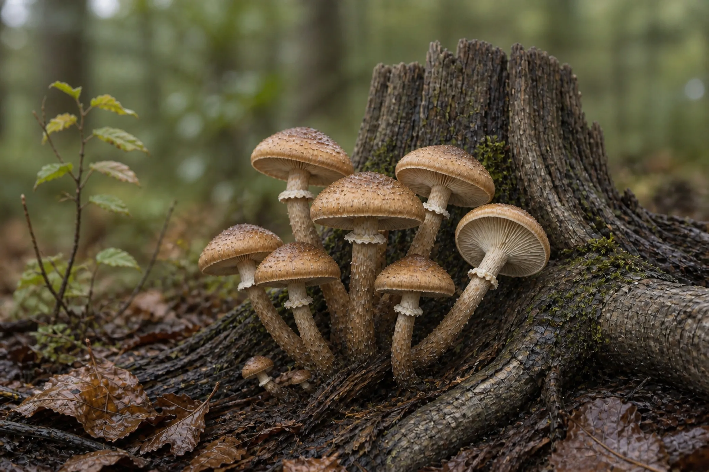
Quick facts
- Scientific name
- Armillaria mellea
- Common names
- Honey fungus
- Taxonomy
- Armillaria · Physalacriaceae
- Edibility context
- Conditional: This database label is context, not remote clearance of a wild specimen; identity, preparation, spoilage, contamination, and individual reaction matter.
- Spore print
- White.
- Toxicity context
- Moderate: Risk includes misidentification and individual reaction. Consult current local toxicology guidance before any food decision.
- Season
- September, October, November
- Growing difficulty
- Not Cultivated
- Regions
- Europe, introduced or related species elsewhere
Safety limit: These labels summarize documented context; they never authenticate a wild specimen. Cooking does not make a poisonous species safe.
Honey fungus is a group name before it is a species name
“Honey fungus” is applied to several species in the genus Armillaria, not only Armillaria mellea. That distinction matters because species distributions, hosts, appearance, and pathogenic behavior differ. The RHS records seven Armillaria species in the United Kingdom and reports A. mellea and A. gallica most often from gardens. A collection elsewhere should be interpreted through a current regional key rather than forced into the European name in this title.
Treat A. mellea as a hypothesis supported by a complete collection record. Fruiting bodies can place a specimen in an Armillaria-like group, but closely related species may require microscopic characters, culture work, mating studies, or molecular testing. Scottish diagnostic guidance is explicit that laboratory PCR can separate regional species that are difficult to distinguish reliably by field characters alone.
Start with the whole cluster and its concealed substrate
Honey fungus often fruits in dense tufts at the base of a tree, around a stump, or apparently from soil above buried roots. Before lifting anything, photograph the cluster’s connection to the host. Probe only where access and tree care allow; a “soil-fruiting” cluster may actually attach to a root several centimeters below the surface. Record whether the wood is living, recently dead, decayed, processed, or unknown.
Fresh caps are commonly honey-yellow to ochre or brown, often darker toward the center, but rain can wash them pale and dry weather can deepen or dull the color. Fine darker scales may be clearer on young caps and become sparse with expansion. Color and scaliness therefore need photographs across several ages, not one weathered cap.
Measure cap width, stem length, and stem thickness on multiple intact fruit bodies. Crowded clusters distort one another, so a single unusually long stem or lopsided cap is not a stable character. Split one representative specimen lengthwise to document solid or fibrous tissue, internal insect damage, and the exact cap-to-stem connection.
Read gills, ring, stem, and spores as a linked set
The gills should be recorded while young and again at maturity. In the broad A. mellea concept they are pale, attached to slightly running down the stem, and may discolor with age. Photograph the gill attachment from the side; an overhead picture cannot preserve this evidence.
A membranous ring on the upper stem is an important clue for A. mellea, but rings tear, collapse, and disappear from old or handled specimens. Document the underside and upper surface of the ring where possible. A ringless cluster should not be “repaired” by assuming weather removed it: ringless Armillaria concepts and unrelated wood-growing fungi change the comparison set.
The stems are typically fibrous and crowded together, sometimes tapering or joining near the base. Record surface texture above and below the ring, basal color, and whether several stems share attached woody material. White spore deposit supports Armillaria, but it is not unique. Make a deposit from more than one mature cap when practical and keep individual caps separated so a mixed cluster cannot produce a misleading combined result.
Rhizomorphs and white mycelial fans are tree-pathology evidence
Black, cord-like rhizomorphs—often described as bootlaces—can travel beneath bark or through soil from colonized roots. White, fan-shaped mycelial sheets may occur between bark and wood near the root collar. These structures are more informative for diagnosing an Armillaria infection than cap color alone, yet neither should be exposed by cutting into a valuable tree without permission and appropriate arboricultural practice.
RHS guidance describes root decay, white fungal growth beneath bark, rhizomorphs, and plant decline as characteristic evidence. Mushrooms are seasonal and short-lived; the perennial infection can remain when no caps are visible. Conversely, mushrooms near a declining tree do not prove that Armillaria initiated the decline. Drought, construction damage, waterlogging, other pathogens, and root disturbance may precede or accompany colonization.
Map the evidence rather than jumping from mushroom to diagnosis: note crown thinning, branch dieback, basal lesions, resin or gum, root-collar condition, rhizomorph position, host species, drainage, recent works, and the location of fruiting. A qualified arborist or plant-pathology service should assess valuable trees and any tree that could strike people or property.
Pathogen, opportunist, and decomposer are not interchangeable labels
An Armillaria individual can persist in woody tissue and extend to new roots, but disease outcome depends on fungus species, host susceptibility, root contacts, site conditions, and plant stress. RHS research distinguishes the strongly pathogenic garden species A. mellea from the generally weaker A. gallica, while noting that stressed plants may still be killed by the latter. That is why species-level diagnosis can materially change management advice.
Do not interpret every stump cluster as an emergency for every nearby tree. Fruiting from dead wood may represent continued decomposition, while rhizomorphs and mycelial fans on living roots support an active disease investigation. Equally, absence of mushrooms is not proof that roots are free of infection. Keep observations tied to the exact host and sampling point.
Separate A. mellea from the most consequential lookalikes
Galerina marginata: rusty-brown spores and a deadly risk
Galerina marginata grows on wood and can possess a ring, making a casual “brown cap plus ring” decision unsafe. Its spore deposit is rusty brown rather than white, and its stature, cap surface, gill maturation, and stem texture differ when intact material is compared. Spore color is a separating character, not permission to eat: a mixed collection can put two species in the same basket or even on the same log.
Hypholoma clusters: darker spores and different gill development
Sulphur tuft and related Hypholoma species also form conspicuous clusters on wood. They lack the same persistent membranous ring, and mature gills reflect their darker spore deposits. Cap color varies enough that “yellow” versus “honey” is a poor first split. Preserve young and mature specimens so gill change is visible.
Other Armillaria: regional keys may outrank photographs
Other honey fungi can overlap in cluster form, pale gills, and white spores. Ring presence, cap ornament, stem characters, host, region, and microscopic or molecular evidence may all matter. A photograph that supports Armillaria may still be insufficient for A. mellea. Record the broader identification honestly when the species boundary is unresolved.
Build an identification record that can be checked later
Use the mushroom photo checklist before separating the cluster. Photograph habitat, host base, entire tuft, cap surface, gills, ring, complete stem, attached wood, and a lengthwise section. Add a ruler and neutral color reference when available. Record date, approximate location, recent rain, nearby trees, and whether fruiting recurs in the same position.
Then use the spore-print method on individually labeled caps. Note actual deposit color under neutral light rather than copying an expected result. If the collection affects a tree-health decision, retain a specimen as directed by the diagnostic service; photographs alone may not support species-level testing.
Food reports do not provide remote clearance
Honey fungus has regional food traditions, but species naming, preparation customs, and individual tolerance vary. Gastrointestinal reactions are reported, and a dangerous wood-growing lookalike can enter a hurried collection. MushroomScope does not recommend eating a specimen identified from this page, an app, or a photograph. Cooking does not neutralize amatoxins in a misidentified Galerina.
If a locally qualified identifier confirms a collection and local guidance permits its use, freshness, legal collection, contamination, and personal health still require separate decisions. Do not use old, waterlogged, insect-damaged, roadside, chemically treated, or mixed material. Do not give a first trial to another person as proof of safety.
After symptoms following any wild mushroom exposure, contact local emergency services or a poison center promptly. Preserve uncooked specimens, cooked leftovers, photographs, and meal timing. Do not wait for a web identification to become certain before seeking help; the poisonous mushroom warning guide explains why symptom-free intervals can matter.
Garden and tree management needs a site diagnosis
Removing visible mushrooms does not remove an underground root and wood infection. Likewise, digging broadly for rhizomorphs can damage healthy roots and spread disturbance. Management depends on which Armillaria is present, which plants are affected, the extent of colonized woody material, site drainage, and the value and structural condition of the host.
For a garden specimen, document affected plants and seek a plant-health diagnosis before major excavation or removal. For mature trees, use a qualified arborist because root decay can create structural risk that crown appearance alone does not reveal. Chemical “cures” and home remedies should not substitute for diagnosis; the RHS reports no available chemical control for garden honey fungus and emphasizes cultural and removal decisions tailored to the site.
Why home cultivation is the wrong objective
This is a root-associated pathogen and wood decomposer, not a sensible beginner food crop. Introducing wild tissue or an unauthenticated culture to logs, beds, or garden soil could move an unwanted organism near susceptible woody plants. A clean-looking culture also does not prove species identity.
Growers seeking clustered culinary mushrooms should use authenticated cultures of established cultivated species and contained, species-specific methods. The beginner growing guide covers that safer workflow. Do not attempt to “rescue” a wild honey-fungus culture into an indoor food project.
Frequently asked questions
Can honey fungus be identified from one photograph?
No. The minimum useful record includes complete fruit bodies, gills, ring, stem base, spore deposit, exact substrate or host, region, and several developmental stages. Closely related Armillaria may require laboratory work.
Does honey fungus always mean a living tree is dying?
No. It can fruit from dead stumps and buried roots, and tree decline can have multiple causes. Rhizomorphs, white mycelial fans, root decay, host condition, and professional site assessment determine the significance.
What color is a honey fungus spore print?
The expected deposit is white. That helps separate some brown-spored wood fungi but cannot by itself confirm A. mellea or clear a collection for food.
Is honey fungus edible after thorough cooking?
Cooking cannot correct a mistaken identity, and even correctly identified honey fungi have variable food traditions and tolerance reports. This page does not provide edibility clearance.
Can honey fungus be cultivated at home?
It should not be treated as a beginner crop. Its root-pathogen ecology and regional species uncertainty make introducing wild or unauthenticated material an unnecessary plant-health risk.
References
- Global Biodiversity Information Facility. Armillaria mellea species record. Taxonomic backbone and occurrence context.
- National Center for Biotechnology Information. NCBI Taxonomy Browser. Independent lineage and nomenclature checkpoint.
- North American Mycological Association. Mushroom Poisoning Syndromes. Poisoning response and syndrome context.
- U.S. Food and Drug Administration. Selecting and Serving Produce Safely. General handling guidance for mushrooms used as food.
- Royal Horticultural Society. Honey fungus. Garden symptoms, rhizomorph biology, regional species, and management context.
- Science & Advice for Scottish Agriculture. Armillaria diagnostics. Regional species separation and PCR diagnostic context.
Editorial review: September 16, 2026. This article has editorial review only and no named mycologist, clinician, or arborist review. Taxonomy, regional range, tree-risk advice, and poison-response contacts should be rechecked at every substantive update.
Source quality notes
MushroomScope cites sources that match the page scope, such as taxonomic databases, extension guidance, food-safety agencies, food-composition databases, and peer-reviewed or institutional health references. Sources support context and uncertainty; they do not turn an online page into specimen identification, medical advice, or a tested recipe record.
Frequently asked questions
Can Honey fungus be identified from one photograph?
No. Identification requires the complete specimen, fertile surface, attachment, substrate, habitat, region, developmental stage, and sometimes microscopy or DNA.
Is wild Honey fungus safe to eat?
This page cannot clear a wild specimen for food. Obtain in-person confirmation from a qualified local identifier and follow local guidance; cooking cannot make a poisonous lookalike safe.
Where does Honey fungus grow?
A root pathogen and wood-decayer fruiting from roots, stumps, and buried wood. Regional records and host associations should be checked before applying this summary to a local collection.
What is the spore print of Honey fungus?
The expected deposit is white, but print color overlaps among fungi and cannot confirm this species by itself.
Can Honey fungus be cultivated at home?
It is not a simple beginner crop; use authenticated commercial material and species-specific professional guidance rather than wild tissue.
Related guides
identification
How to Identify Mushrooms: A Beginner's Field Guide
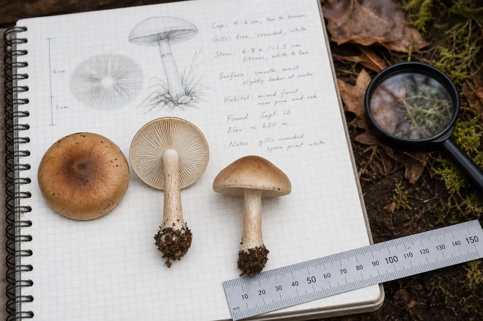
Build an identification from the whole specimen, substrate, habitat, spore deposit, and location—not from a single photograph.
identification
Mushroom Identification Safety Checklist
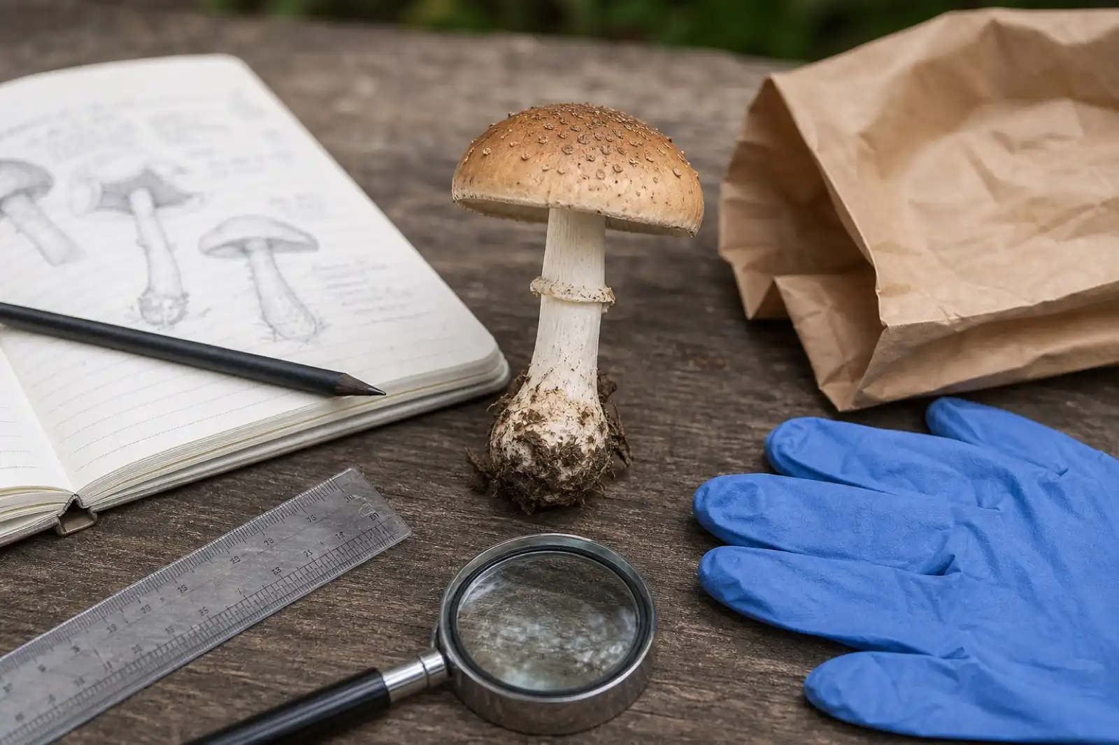
Use a conservative checklist before handling, documenting, or discussing any wild mushroom, especially when edibility is being considered.
mushrooms
Cantharellus cibarius: Chanterelle Identification Guide
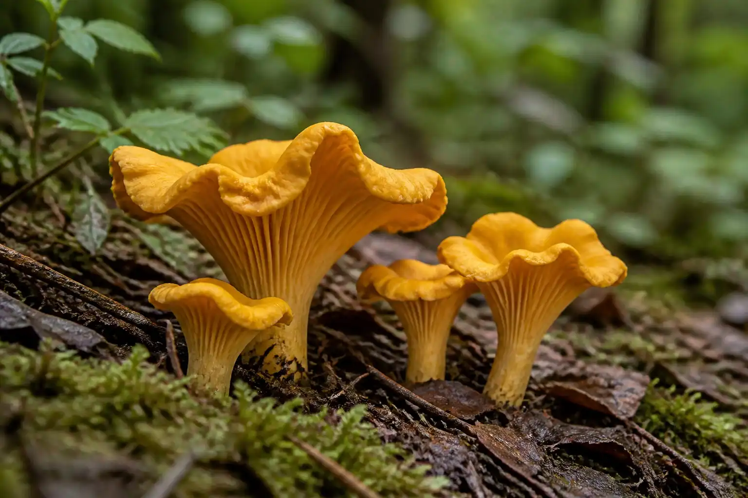
Learn chanterelle identification, false gills, habitat, season, poisonous lookalikes, edibility, nutrition, and why wild finds need local verification.
Continue exploring
Browse more practical guides in Mushroom Encyclopedia or visit the mushroom encyclopedia.